Modern regenerative dentistry increasingly relies on autologous blood concentrates to improve healing, reduce complications, and enhance treatment predictability. Today, PRP for dental procedures and platelet-rich fibrin (PRF) are widely used in oral surgery, implantology, and periodontics to support tissue regeneration, improve surgical outcomes, and shorten recovery times.
While clinicians often focus on centrifugation protocols and preparation techniques, the collection tube itself plays an equally important role in determining the quality of the final product. Tube composition, additives, and centrifugation parameters directly influence platelet concentration, fibrin architecture, growth factor release, and ultimately clinical outcomes.
This article explores how PRP for dental applications and PRF contribute to extraction healing, implant osseointegration, sinus augmentation, bone grafting, and soft tissue regeneration—and why selecting the appropriate tube can significantly improve treatment outcomes.
Understanding the Difference Between PRP and PRF in Dentistry
Although both PRP and PRF originate from the patient’s own blood, they differ substantially in structure, preparation method, and biological behavior.
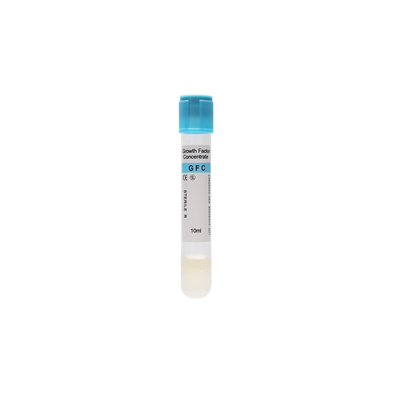
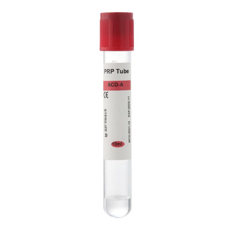
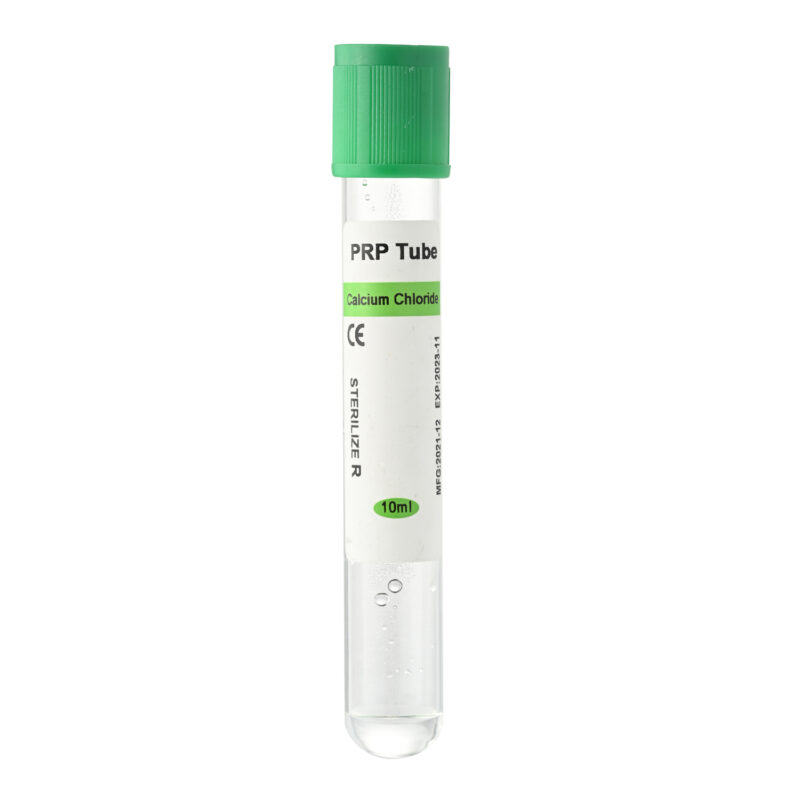
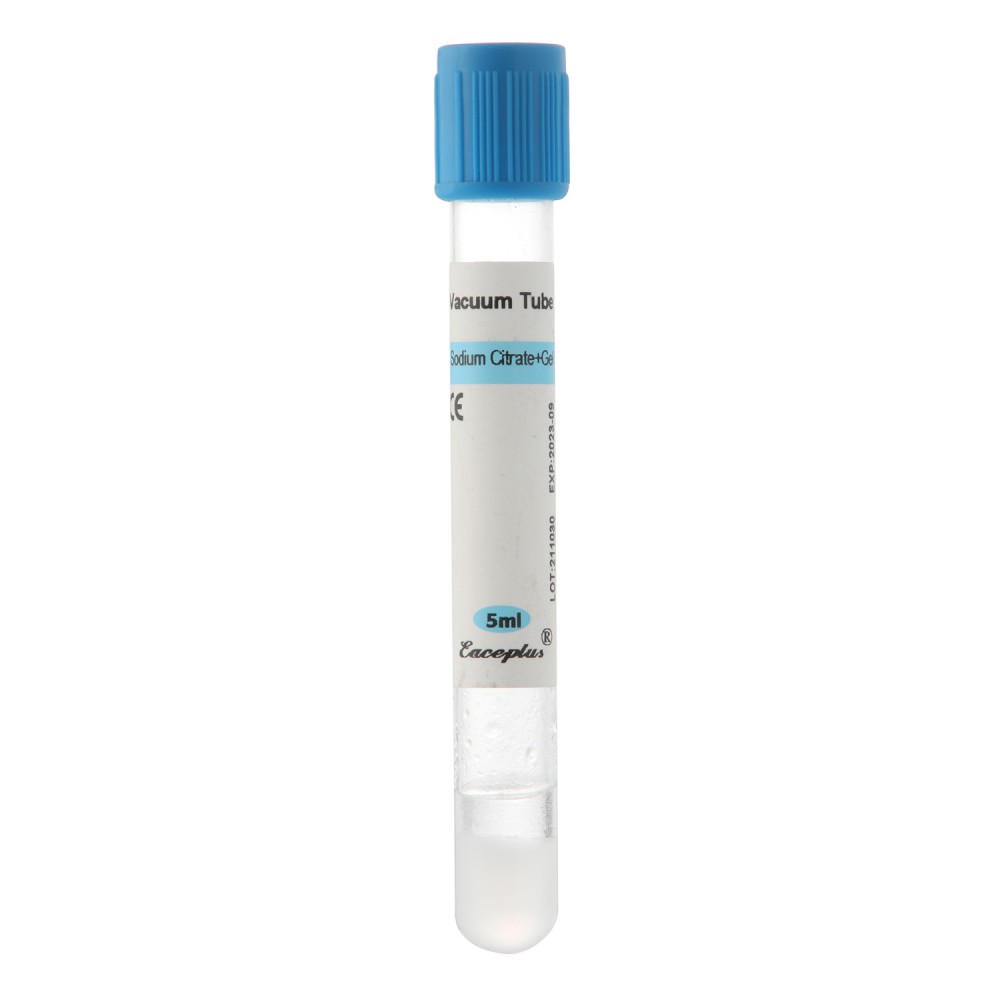
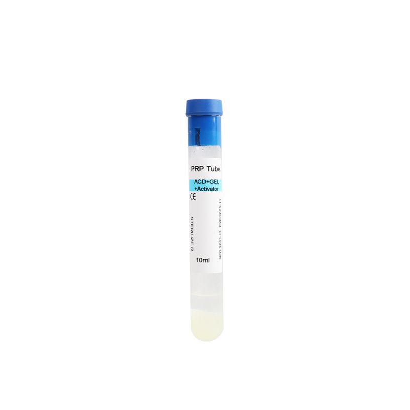
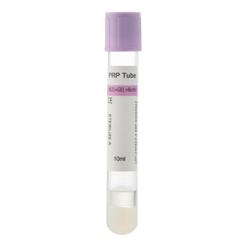
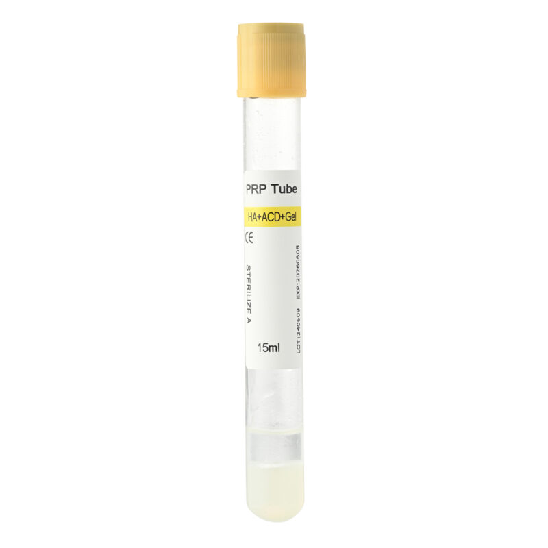
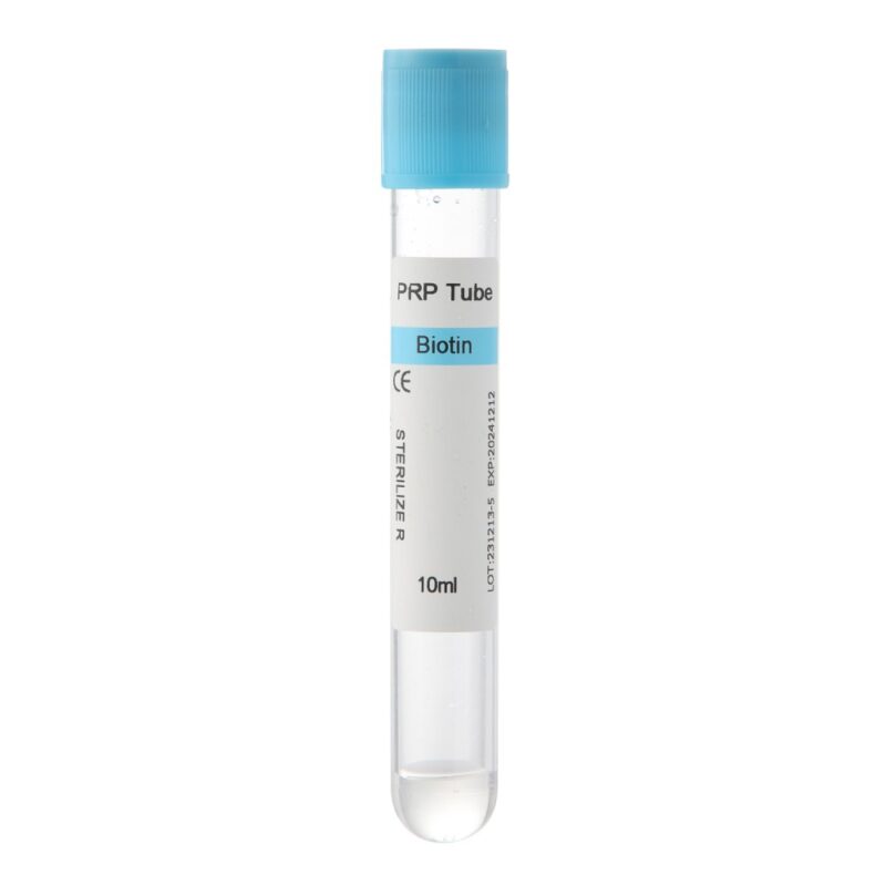
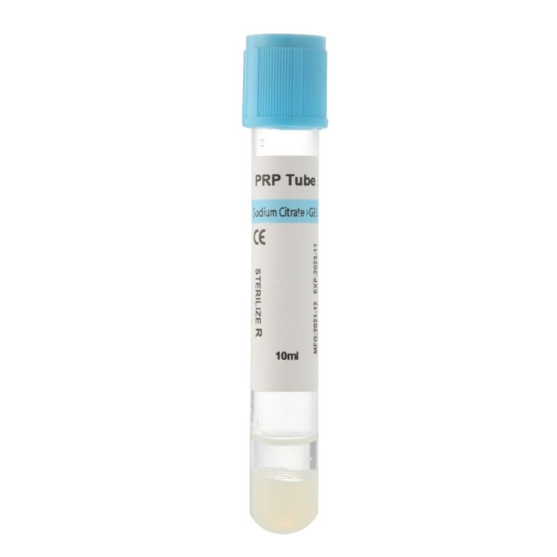
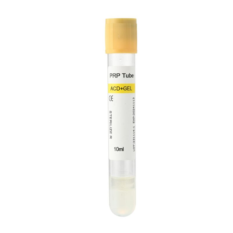
PRP (Platelet-Rich Plasma) is a liquid concentrate containing elevated platelet levels suspended in plasma. To prepare PRP, clinicians use an anticoagulant such as ACD-A or sodium citrate during blood collection to prevent immediate clotting. Before clinical application, PRP typically requires activation using agents such as calcium chloride (CaCl₂) or thrombin. Once activated, platelets rapidly release a high concentration of growth factors, including PDGF, VEGF, TGF-β, and IGF-1, creating an immediate biological stimulus that supports tissue repair and cellular recruitment.
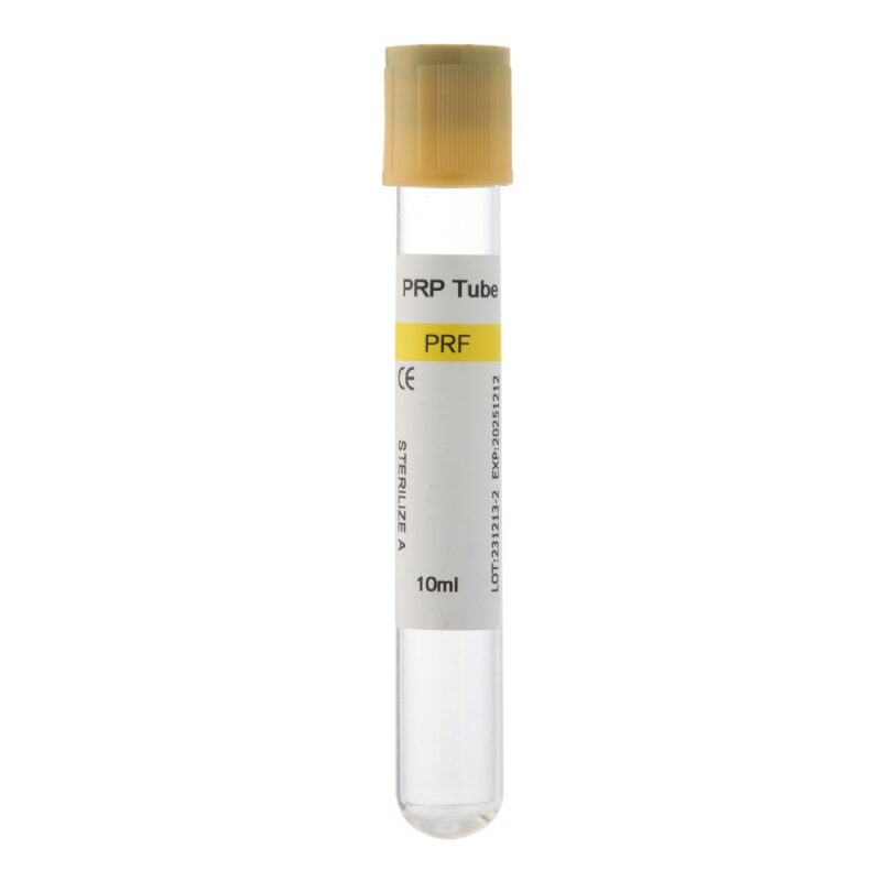
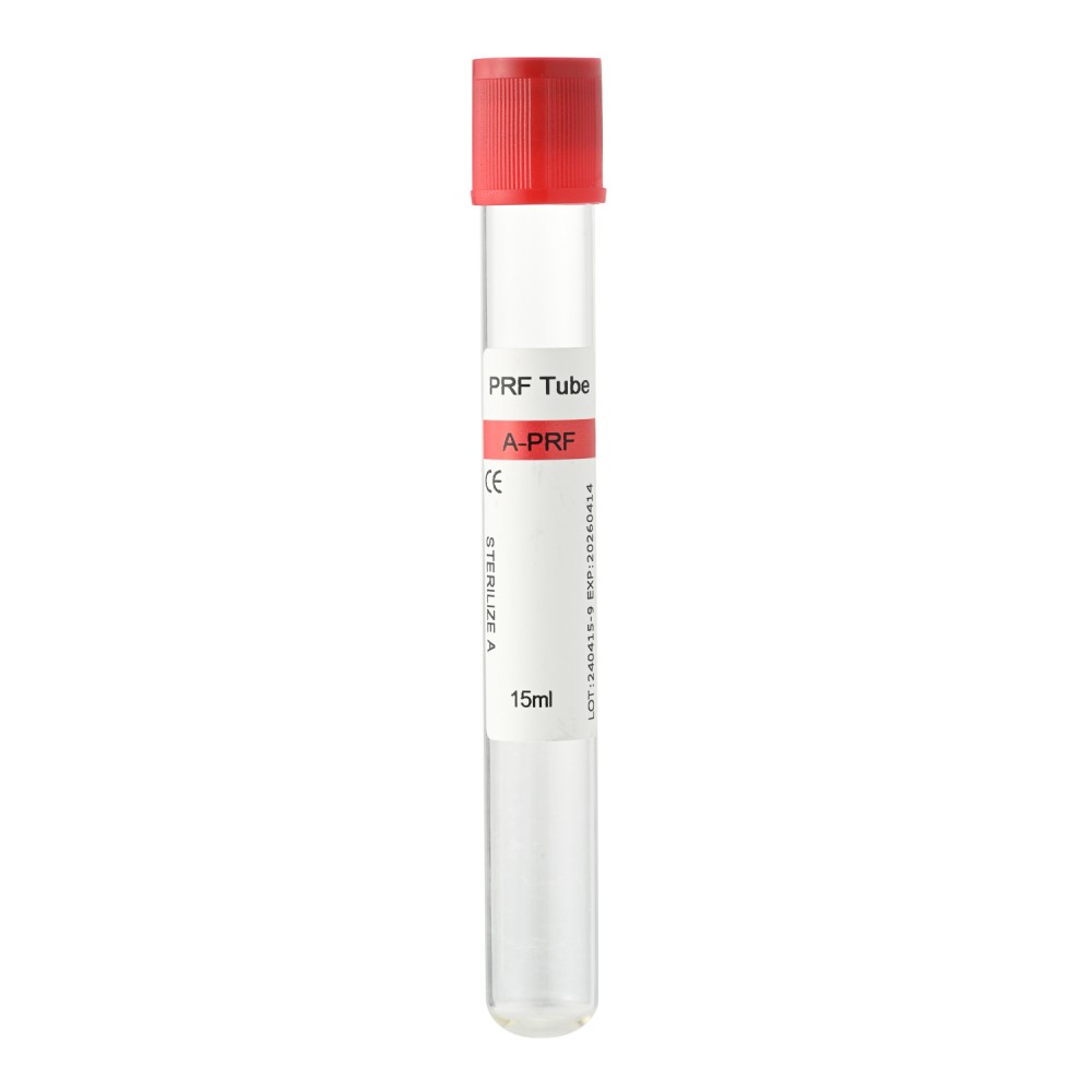
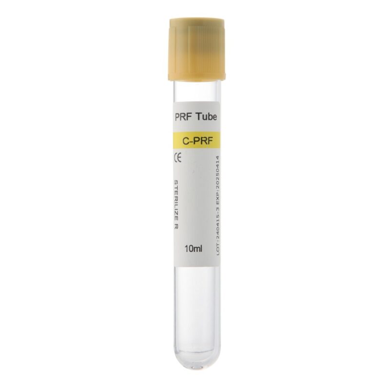
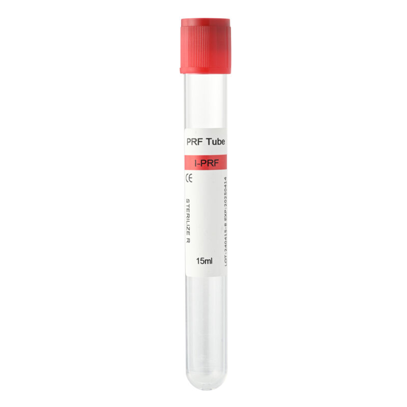
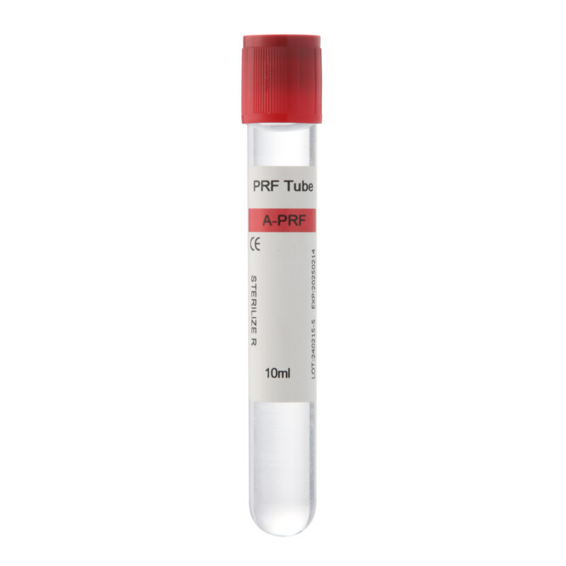
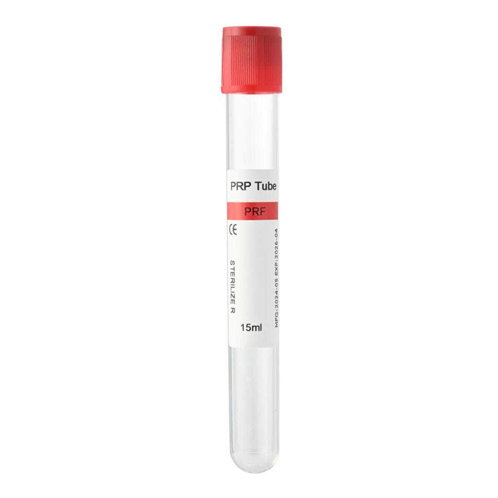
PRF (Platelet-Rich Fibrin) is a completely autologous system prepared without anticoagulants or chemical activators. Natural coagulation begins immediately after blood collection, resulting in a three-dimensional fibrin matrix that traps platelets, leukocytes, and cytokines. Depending on the centrifugation protocol, PRF can be prepared as a solid fibrin clot or membrane (L-PRF, A-PRF) or as a temporary liquid concentrate (i-PRF) that remains injectable for several minutes before naturally polymerizing.
Unlike PRP, which delivers a rapid growth factor burst, PRF provides a gradual and sustained release of regenerative molecules over an extended period, supporting ongoing tissue healing and remodeling.
Biological and Physical Comparison
| Feature | PRP (Platelet-Rich Plasma) | PRF (Platelet-Rich Fibrin) |
|---|---|---|
| Physical Form | Liquid (requires activation) | Solid membrane/clot or injectable liquid (i-PRF) |
| Growth Factor Release | Rapid burst | Sustained release |
| Anticoagulant Required | Yes | No |
| Chemical Activation | Usually required | Not required |
| Scaffold Function | Limited | Excellent |
| Common Dental Applications | Injections, graft hydration, and implant surface treatment | Membranes, socket filling, graft stabilization, sticky bone |
For many regenerative procedures, clinicians utilize both PRP and PRF because they address different biological requirements during healing.
PRP for Dental Extractions and Clot Protection
Following tooth extraction, maintaining a stable blood clot is critical for uneventful healing. Clot disruption may lead to delayed tissue repair, alveolar bone loss, or painful complications such as alveolar osteitis (dry socket).
PRP for dental extractions provides a concentrated burst of growth factors immediately after surgery. When applied to extraction sites, PRP can stimulate angiogenesis, accelerate epithelialization, and support early soft tissue closure.
PRF offers a different advantage. A PRF membrane functions as a biologic dressing that physically protects the extraction socket while simultaneously releasing regenerative signals. Because the fibrin network provides structural stability, PRF is often preferred when clinicians require both clot protection and tissue scaffolding.
For routine extractions, PRP may accelerate initial healing. For surgical extractions, impacted third molars, and compromised healing situations, PRF membranes frequently provide additional benefits due to their mechanical and biological properties.
A dedicated socket preservation tube designed specifically for PRF preparation helps produce a dense and stable fibrin clot suitable for placement within extraction sockets.

PRP Dental Implant Procedures and Osseointegration
Successful implant treatment depends on rapid and predictable osseointegration. Platelets play a significant role during the early healing phase by releasing signaling molecules that recruit osteoblasts and stimulate vascular development around the implant surface.
Today, PRP dental implant protocols are widely used to enhance early healing and improve regenerative outcomes. Liquid PRP is commonly applied to implant surfaces or mixed with particulate graft materials before placement. Once activated, it delivers a concentrated burst of growth factors that support early cellular activity and vascularization around the implant.
Conversely, liquid PRF (i-PRF) has gained widespread popularity for implant surface conditioning. Because it is entirely autologous and free of anticoagulants, i-PRF naturally polymerizes directly onto titanium surfaces, creating a biologically active layer rich in platelets and leukocytes.
Solid PRF membranes can also be placed around implant collars or osteotomy sites to provide sustained growth factor release during the critical early stages of bone formation. Many implantologists combine PRP and PRF to leverage both immediate biologic stimulation and long-term regenerative support.
Sinus Lift and Bone Grafting Applications: The Sticky Bone Concept
Sinus augmentation and bone grafting procedures require predictable vascularization, graft stabilization, and long-term regenerative support.
Historically, clinicians used PRP to hydrate and bind bone graft materials. While effective, PRP primarily provides a short-term growth factor release.
The introduction of injectable PRF (i-PRF) has significantly expanded regenerative possibilities through the creation of “sticky bone.” By mixing particulate bone graft materials—including allografts, xenografts, and synthetic substitutes—with liquid i-PRF, clinicians create a cohesive graft mass that naturally polymerizes within minutes without chemical additives.
This sticky bone complex offers several advantages:
- Improved graft stability
- Reduced particle migration
- Easier handling during surgery
- Sustained release of growth factors
- Enhanced angiogenesis and tissue integration
PRF membranes also play an important role in sinus lift procedures. Clinicians frequently use them to:
- Protect or reinforce the Schneiderian membrane
- Cover lateral window osteotomies
- Improve vascularization within grafted regions
- Act as biologic barriers against soft tissue invasion
Because fibrin quality directly influences membrane handling, elasticity, and regenerative potential, selecting a dedicated dental PRF tube becomes particularly important during extensive augmentation procedures.
Gum Grafting and Soft Tissue Regeneration
Periodontal plastic surgery and mucogingival procedures frequently require soft tissue grafting to increase keratinized tissue, cover exposed roots, and improve esthetic outcomes.
PRP can be injected into recipient sites or applied to connective tissue grafts to enhance early healing. The rapid release of growth factors may reduce postoperative inflammation and stimulate fibroblast activity during the initial healing phase.
PRF membranes provide additional advantages because they combine biologic stimulation with physical tissue support. Clinicians can position PRF membranes beneath coronally advanced flaps or alongside connective tissue grafts to create a natural scaffold that promotes angiogenesis, fibroblast migration, and soft tissue maturation.
Many periodontists now incorporate PRF into recession coverage and soft tissue augmentation procedures because it can improve wound stability, enhance patient comfort, and reduce donor-site morbidity.
Tube Selection and the Controversy Surrounding Silica-Coated Tubes
The success of PRP and PRF therapy depends heavily on the blood collection tubes used during preparation. However, one aspect of tube selection continues to generate discussion within the regenerative dentistry community: the use of silica-coated diagnostic tubes.
Many standard plastic blood collection tubes designed for laboratory diagnostics contain silica-based clot activators. These coatings help serum tubes clot rapidly for laboratory testing, but were not originally developed for regenerative surgical applications.
Several peer-reviewed studies have reported that silica microparticles from clot activator tubes may become incorporated into PRF matrices during centrifugation. Laboratory investigations have suggested that these particles may negatively affect cell viability under certain experimental conditions.
For this reason, many clinicians prefer using:
- Additive-free glass tubes that support natural coagulation without releasing particulate clot activators.
- Medical-grade titanium tubes that promote fibrin formation through surface interactions rather than chemical coatings.
- Specialized silica-free PRF tubes have been developed specifically for regenerative procedures.
Selecting a tube designed for PRF preparation helps minimize unwanted variables and contributes to more consistent fibrin architecture and biologic performance.
How Centrifugation Settings Affect Fibrin Architecture
One of the most overlooked factors in PRP and PRF preparation is the centrifugation protocol.
The speed and duration of centrifugation significantly influence platelet recovery, leukocyte distribution, and fibrin structure.
Higher relative centrifugal force (RCF) generally produces denser fibrin networks but may compress cellular components and reduce biologic activity by concentrating regenerative cells at the bottom of the tube.
Conversely, low-RCF protocols have gained popularity in regenerative dentistry because they help preserve higher concentrations of platelets and leukocytes throughout the fibrin matrix. These protocols may contribute to:
- Improved cellular retention
- Greater growth factor availability
- Enhanced vascularization potential
- Better membrane flexibility
- Improved surgical handling characteristics
Tube design also affects separation quality. Factors such as tube material, internal surface characteristics, vacuum consistency, and additive composition influence clot formation and platelet capture efficiency.
Clinicians should therefore view centrifugation settings and tube selection as a combined system rather than independent variables. Optimizing both factors helps produce blood concentrates that match the biological requirements of specific dental procedures.
Selecting the Right PRP and PRF Tube for Dental Regeneration
Whether performing routine extractions, implant placement, sinus augmentation, bone grafting, or periodontal soft tissue procedures, clinicians should view tube selection as a critical component of regenerative success.
The right tube supports optimal platelet recovery, predictable fibrin formation, and consistent growth factor delivery. Combined with appropriate centrifugation protocols, a well-designed PRP or PRF tube can improve handling characteristics and enhance biologic performance across a wide range of dental applications.
Siny PRP offers specialized dental PRF tubes, additive-free glass tubes, and regenerative blood collection solutions designed to meet the needs of oral surgeons, implantologists, and periodontists. If you are evaluating PRP or PRF preparation systems for your practice, our team can help identify the most suitable solution for your clinical workflow.
FAQs
Is PRP or PRF better for dental implants?
Neither is universally superior because they serve different functions. PRP provides an immediate growth factor burst that supports early healing, while PRF offers sustained growth factor release and scaffold support. Many clinicians combine both approaches to maximize regenerative benefits.
Why do PRF tubes not contain anticoagulants?
PRF relies on the natural coagulation cascade to form a three-dimensional fibrin matrix. Anticoagulants would prevent the clot formation necessary for producing PRF membranes and fibrin-rich grafting materials.
Why are silica-coated diagnostic tubes controversial for PRF preparation?
Several studies have reported that silica microparticles from clot activator tubes may become incorporated into PRF matrices during centrifugation and may negatively affect cell viability in vitro. As a result, many clinicians prefer additive-free glass tubes, titanium tubes, or silica-free PRF tubes specifically developed for regenerative applications.
How long do PRF membranes release growth factors?
Research indicates that PRF matrices can provide a sustained release of growth factors and cytokines for approximately 10 to 14 days, although the exact duration may vary depending on preparation protocols and fibrin architecture.
Does centrifugation speed affect PRF quality?
Yes. Centrifugation speed and relative centrifugal force directly influence fibrin architecture, platelet retention, leukocyte concentration, membrane density, and overall regenerative potential.
What is sticky bone and how is it made?
Sticky bone is a cohesive bone graft complex created by mixing particulate bone graft materials with liquid i-PRF. The mixture naturally polymerizes within minutes, producing a biologically active graft matrix that improves handling, reduces graft migration, and supports tissue regeneration.