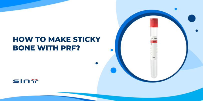
If you work with dental implants or regenerative dentistry, you may have heard the term “sticky bone.” But how to make sticky bone with PRF, and why is injectable platelet-rich fibrin (i-PRF) often used for this purpose?
In simple terms, sticky bone is prepared by collecting blood, producing liquid i-PRF through centrifugation, mixing it with particulate bone graft material, and allowing a fibrin network to develop. As the fibrin forms, it binds the individual graft particles into a more cohesive and moldable mass.
This change can make particulate graft material easier to transfer, shape, and stabilize during placement. At the same time, i-PRF introduces autologous fibrin, platelets, leukocytes, and signaling molecules associated with the healing process.
However, successful sticky bone preparation involves more than mixing liquid PRF with bone particles. Blood collection, centrifugation, timing, graft characteristics, and the PRF preparation system all affect the workflow.
This guide explains how i-PRF sticky bone is prepared, why the graft becomes cohesive, where the technique is used in dentistry, and which preparation mistakes professionals should avoid.
What Is Sticky Bone and Why Is i-PRF Used?
Particulate bone graft materials are widely used in implant dentistry and oral bone regeneration. Depending on the procedure, clinicians may choose autogenous bone, allografts, xenografts, synthetic substitutes, or a combination of materials.
Particulate grafts adapt well to irregular defects. However, loose particles can move or scatter during handling. They may also be harder to shape into the desired contour before stabilization.
Sticky bone changes these handling characteristics.
When freshly prepared liquid i-PRF is mixed with particulate graft material, fibrinogen in the liquid phase gradually polymerizes into fibrin. This developing fibrin network surrounds and connects the graft particles.
As a result, the mixture becomes more cohesive. Instead of handling many individual particles, the clinician can work with a graft mass that tends to remain together during gentle manipulation.
Why Use i-PRF for Sticky Bone Preparation?
i-PRF is useful because it remains liquid for a limited period after centrifugation. During this working window, it can be collected and mixed with particulate graft material.
Fibrin polymerization then continues naturally.
This creates the characteristic structure of i-PRF sticky bone: bone graft particles distributed within an autologous fibrin matrix.
The potential value of i-PRF is not limited to physical binding. Platelets and other biological components within the preparation are associated with signaling processes involved in tissue repair, angiogenesis, cell migration, and extracellular matrix formation.
For this reason, PRF-based preparations have been studied across several areas of oral and periodontal regeneration.
Still, sticky bone should not be treated as a shortcut around established surgical principles. Defect morphology, vascular supply, graft selection, space maintenance, mechanical stability, membrane use, flap management, and patient-related factors remain important.
How to Make Sticky Bone with PRF: Step-by-Step Guide
The basic workflow is:
Blood collection → i-PRF preparation → liquid i-PRF collection → bone graft mixing → fibrin polymerization → graft placement.
Although the sequence is straightforward, each stage should follow a validated professional protocol.
Step 1: Prepare the Particulate Bone Graft
Prepare the required bone graft material before drawing blood.
The graft may consist of autogenous particles or an appropriate bone substitute selected for the planned regenerative procedure. Particle size, material composition, porosity, and absorption characteristics can affect how the graft interacts with liquid i-PRF.
Place the required amount in a sterile container and have it ready for mixing.
This small preparation step is important because liquid i-PRF has a limited working period. Once it has been collected, the clinical team should be ready to use it without unnecessary delay.
Step 2: Collect the Blood
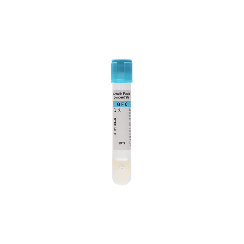
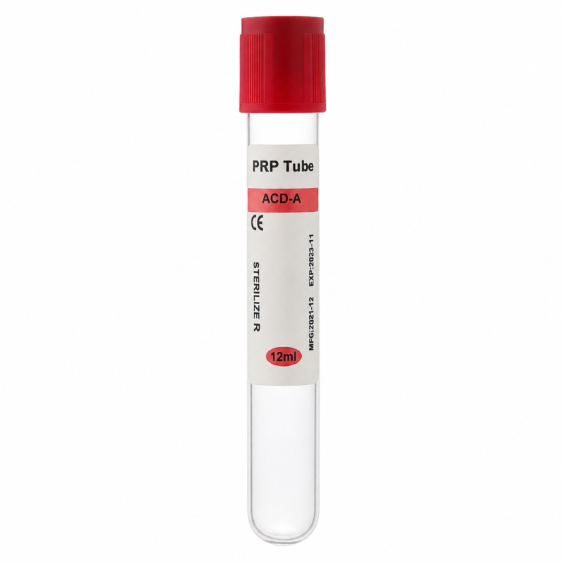
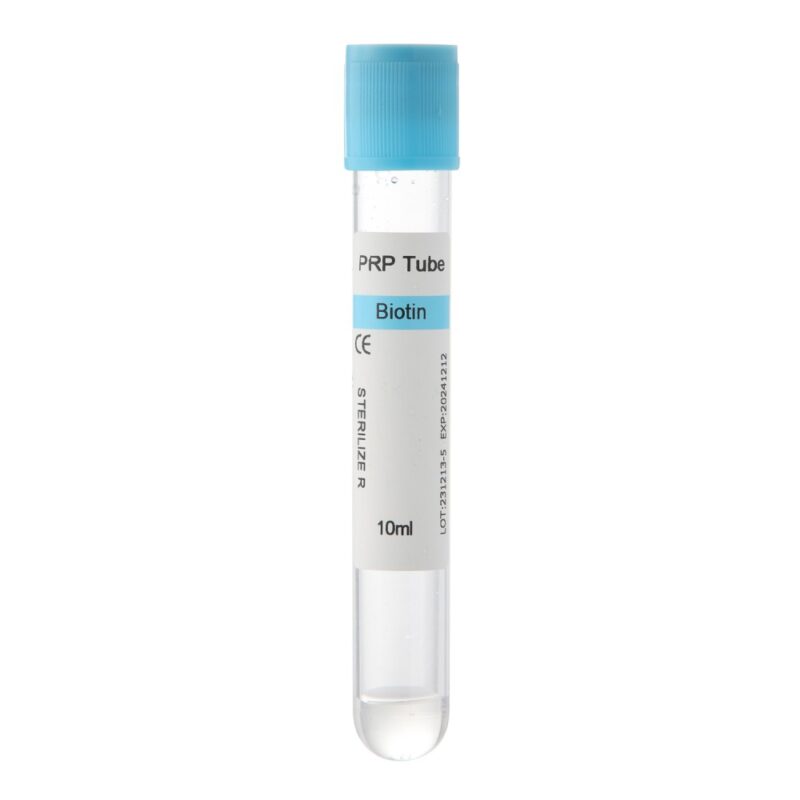
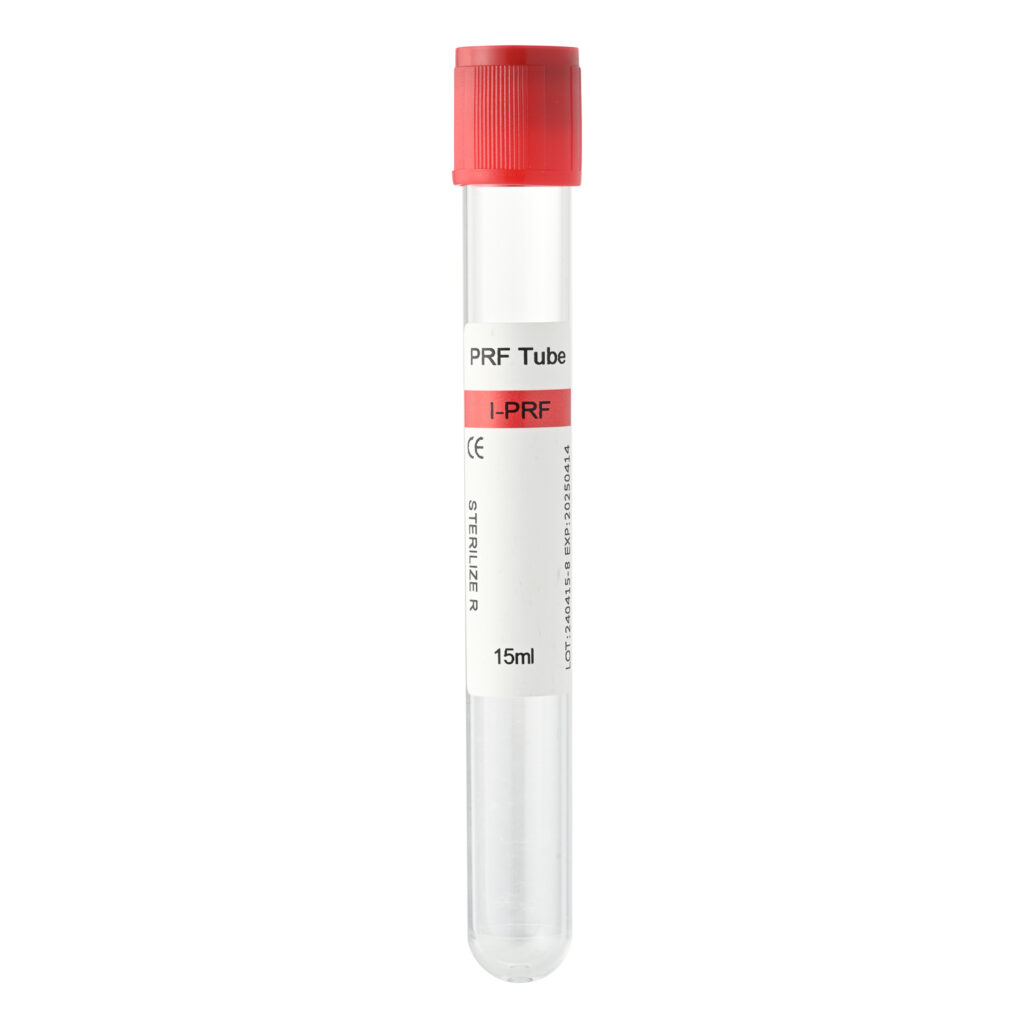
Collect venous blood using a tube suitable for the intended i-PRF preparation protocol.
The collection tube is one component of the overall preparation system. Tube characteristics, blood volume, centrifuge compatibility, rotor design, centrifugal force, processing time, and handling protocol should be considered together.
The centrifuge and graft materials should already be ready before blood collection begins.
Efficient handling matters because coagulation starts naturally after blood collection. An unnecessary delay before centrifugation can change the characteristics of the final PRF preparation.
Step 3: Centrifuge the Blood to Prepare i-PRF
Place the collected tubes into the centrifuge according to the validated i-PRF protocol.
Be careful when copying centrifugation parameters from another system.
RPM alone does not define the centrifugal force applied to the sample because rotor radius also affects relative centrifugal force (RCF). Therefore, a protocol developed for one centrifuge should not automatically be transferred to another machine based only on RPM.
The objective is to obtain a liquid PRF fraction that remains suitable for collection and immediate mixing with the graft.
For that reason, clinicians should follow the centrifugation protocol validated for their tube and centrifuge system rather than assuming that one universal RPM and time combination works for every setup.
Step 4: Collect the Liquid i-PRF
After centrifugation, collect the liquid i-PRF fraction according to the established clinical protocol.
Timing becomes especially important at this stage.
Unlike a solid PRF membrane, i-PRF is collected while it is still fluid enough to mix with another biomaterial. Once collected, fibrin formation continues.
Therefore, move directly to graft mixing rather than leaving the liquid i-PRF standing unnecessarily.
Step 5: Mix i-PRF with the Bone Graft
Transfer the freshly prepared liquid i-PRF to the particulate bone graft.
Gently mix the two components so that the graft particles become evenly incorporated into the liquid phase. The aim is not simply to soak the bone substitute. Instead, the particles should become distributed throughout the developing fibrin matrix.
The amount of liquid required can vary.
Particle size, graft composition, absorption characteristics, defect size, and the desired handling consistency all influence the mixture. This is why a single liquid-to-graft ratio should not automatically be applied to every clinical situation.
At first, the graft may still appear relatively loose or wet. Its handling characteristics change as fibrin polymerization progresses.
Step 6: Allow the Fibrin Network to Form
After mixing, allow the fibrin matrix to develop.
As polymerization progresses, the graft generally becomes more cohesive and moldable. Individual particles begin to remain together during gentle handling rather than immediately separating.
This transition creates the characteristic “sticky” behavior.
There is no universal waiting time for every preparation system. Polymerization can vary with the PRF protocol, graft material, handling conditions, and preparation workflow.
Instead of relying only on the clock, the clinician should evaluate the actual handling characteristics of the graft according to the established clinical protocol.
Step 7: Shape and Place the Sticky Bone
Once the desired cohesion has developed, the sticky bone can be shaped according to the defect and treatment plan.
This is where the technique offers a practical handling advantage. A cohesive graft mass can be easier to transfer and contour than dry, loose particles.
However, cohesion does not equal rigid fixation.
The need for a membrane, fixation device, space-maintaining technique, or other regenerative approach depends on the defect. These decisions remain part of the clinician’s surgical plan.
When learning how to make sticky bone with PRF, it is therefore important to look at the complete workflow. Good preparation begins before the graft and i-PRF ever meet.
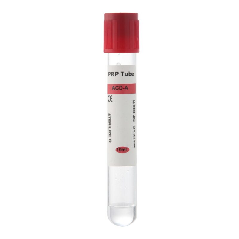
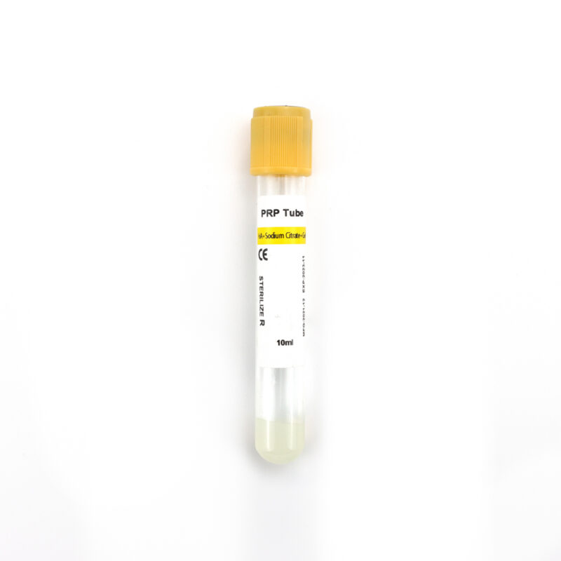
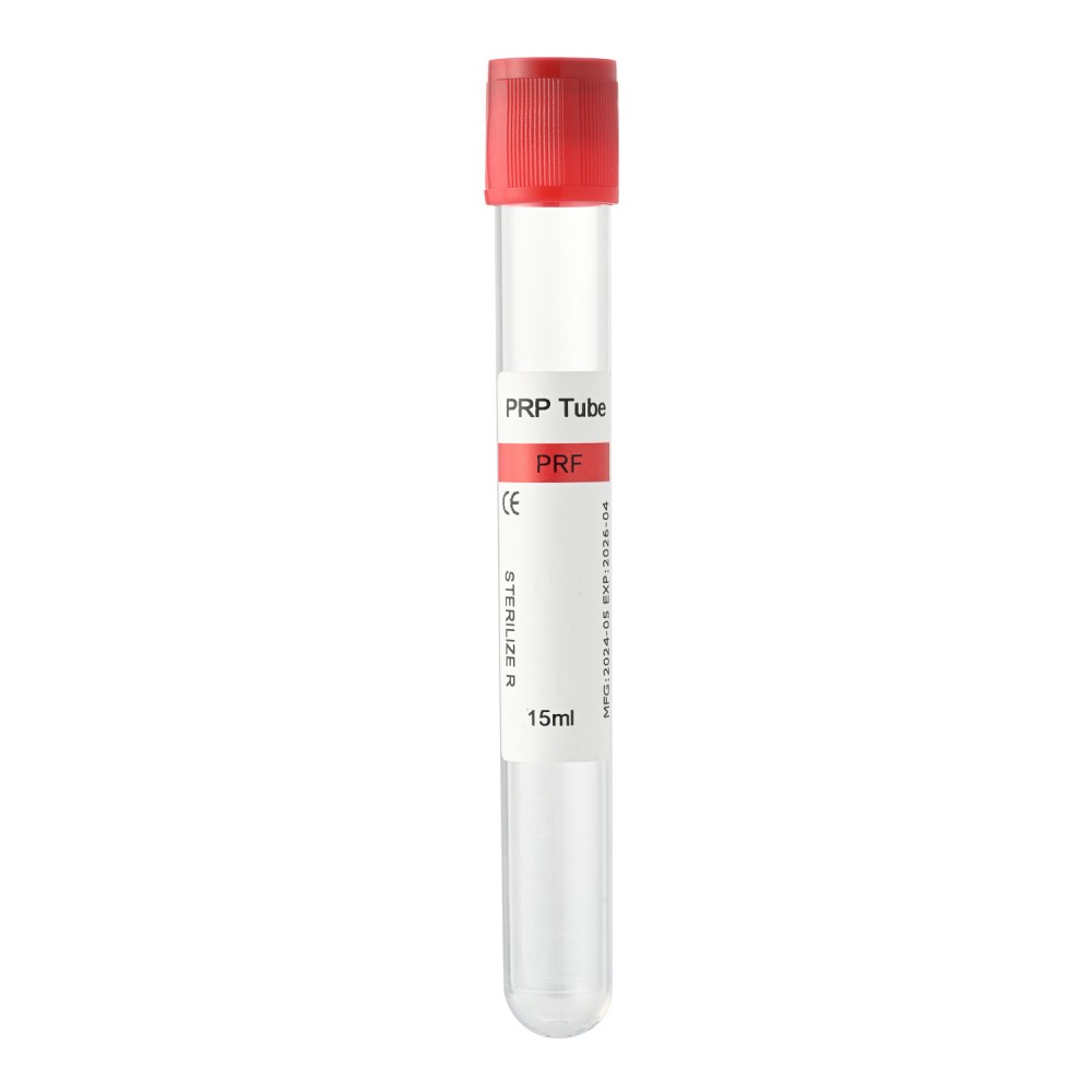
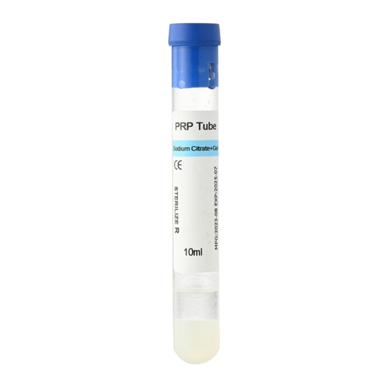
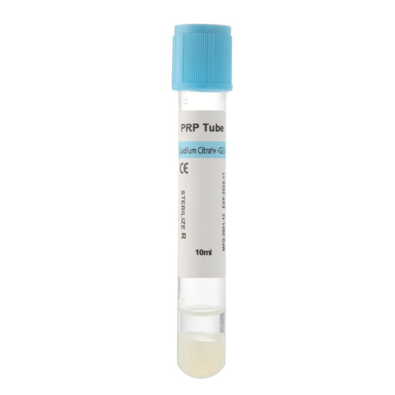
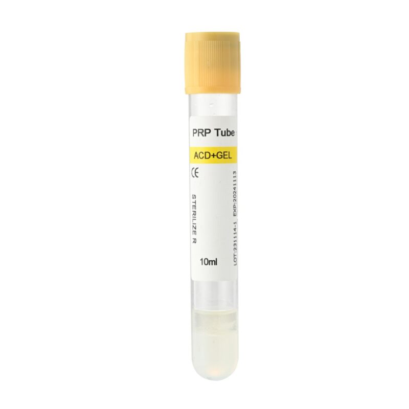
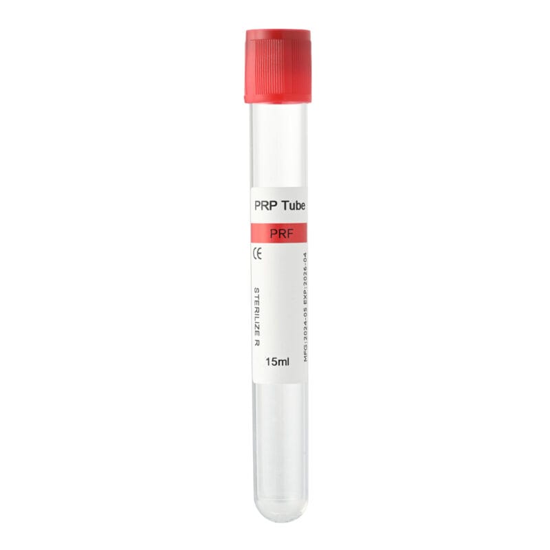
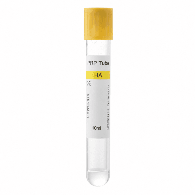
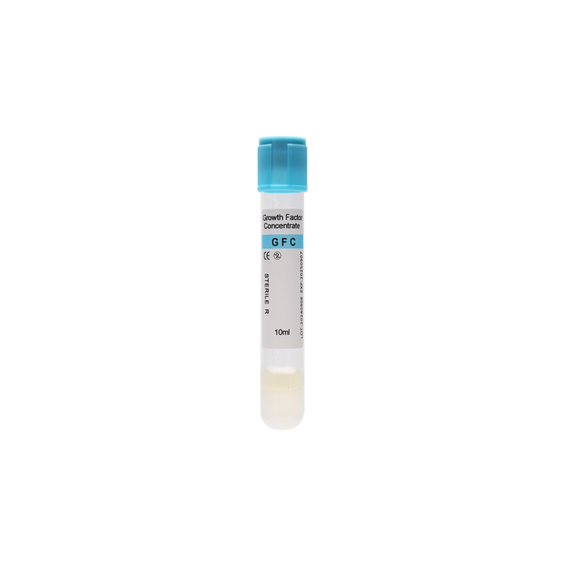
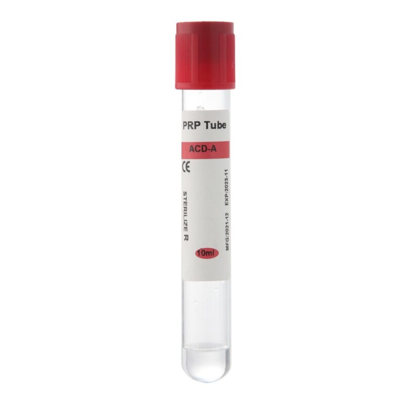
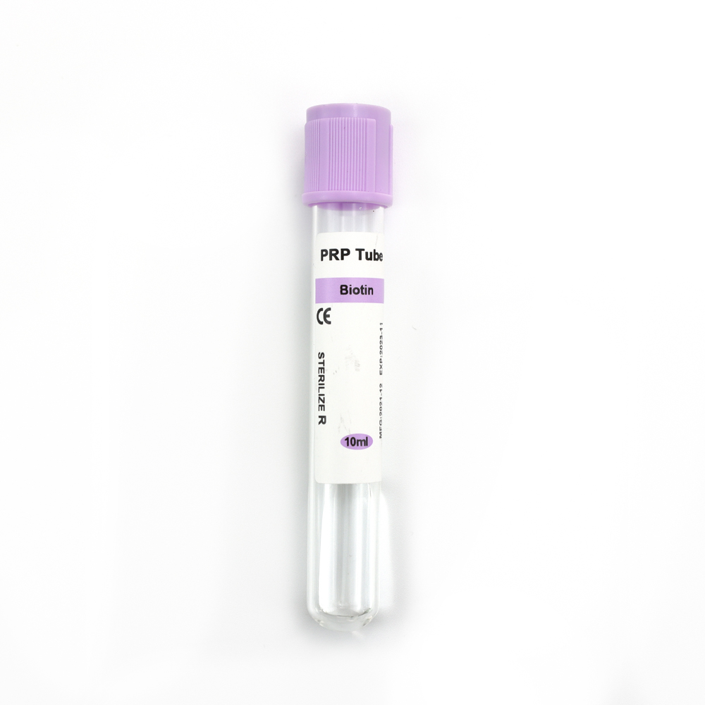
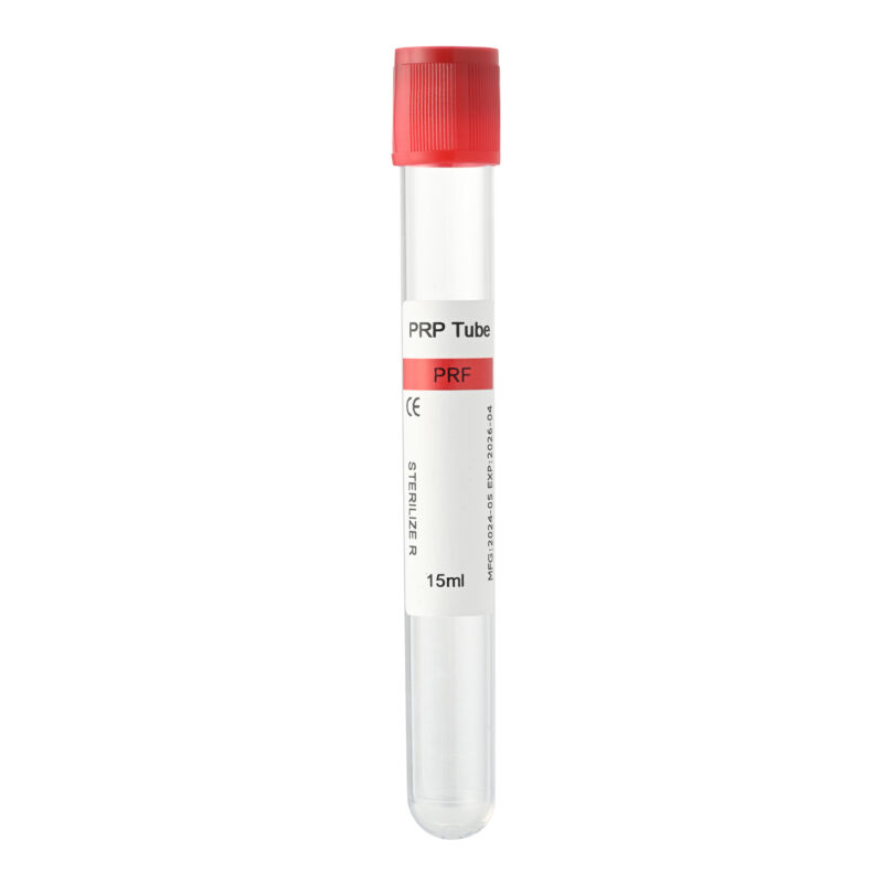
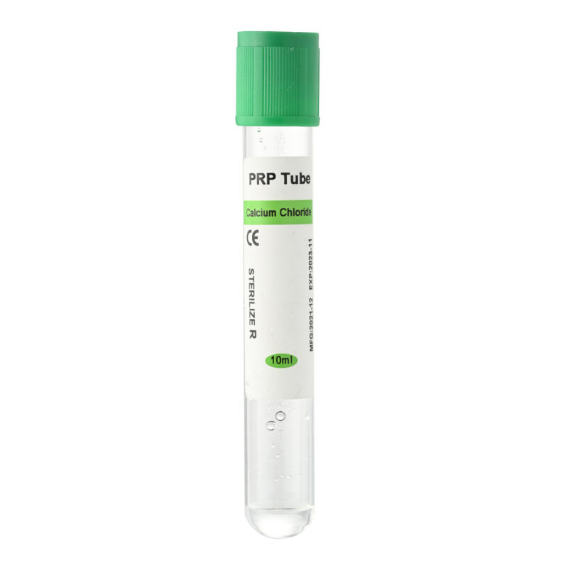
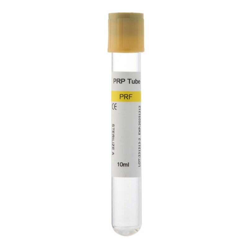
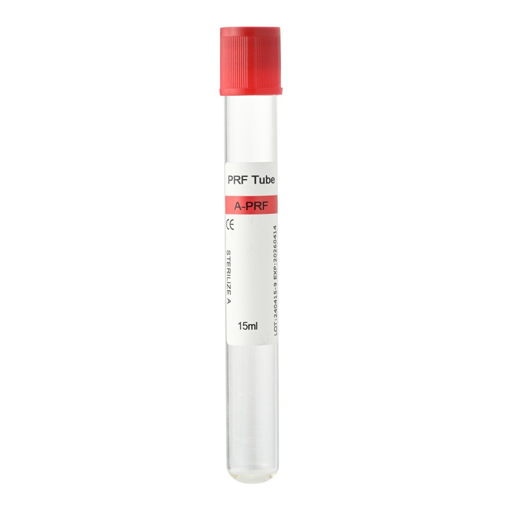
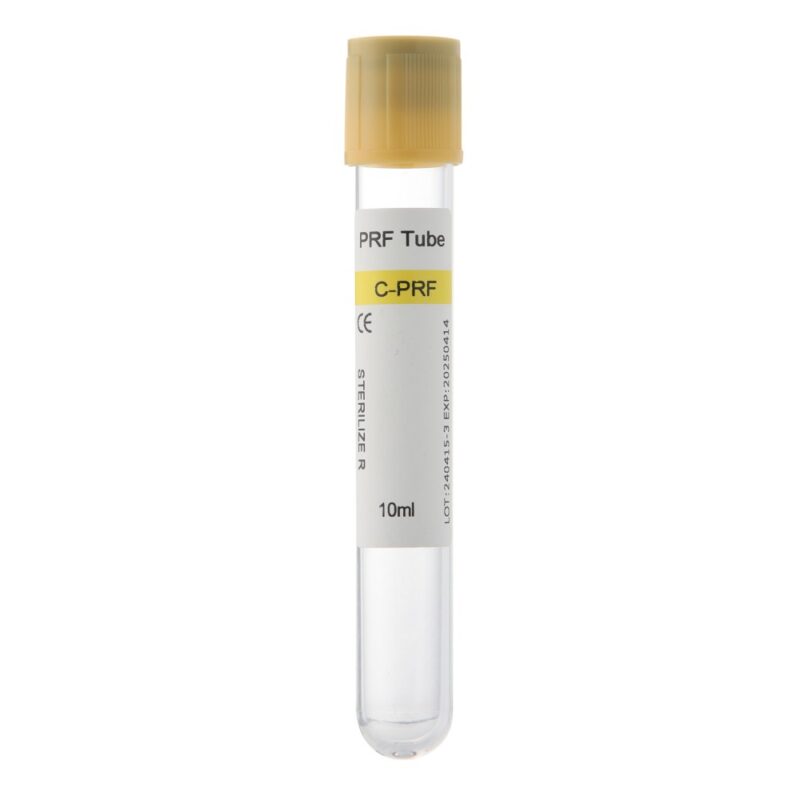
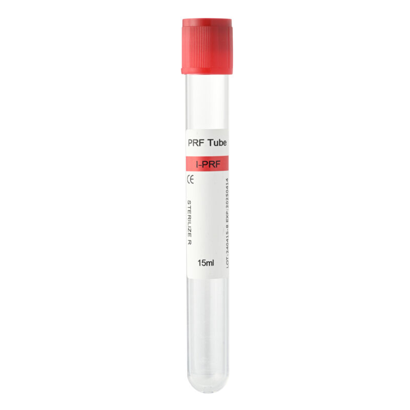
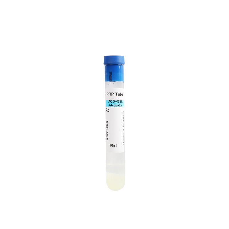
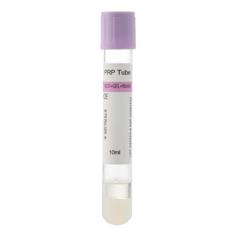
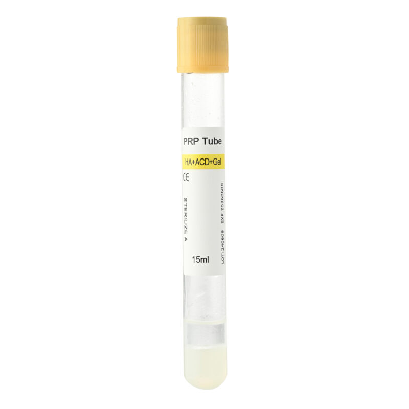
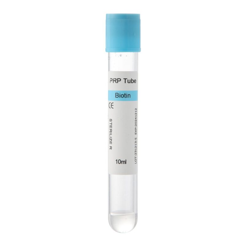
For practices developing an i-PRF workflow, Siny PRP offers PRF tube options that can be explored alongside the clinic’s established centrifugation and preparation protocol.
Why Does i-PRF Make Bone Graft “Sticky”?
The visible difference is easy to recognize: loose graft particles start behaving like a single, manageable mass.
The reason lies mainly in fibrin.
Fibrin Forms a Natural Binding Matrix
During coagulation, fibrinogen is converted into fibrin. The developing fibrin strands form a three-dimensional network around and between the graft particles.
This network acts as a natural binder.
As it develops, particles that previously moved independently become incorporated into the same matrix. This gives the graft greater cohesion during handling.
Importantly, the material does not become a rigid block. It remains adaptable enough to contour to the surgical site.
i-PRF Adds Autologous Biological Components
i-PRF is prepared from the patient’s own blood. Its fibrin matrix contains platelets and other cellular and molecular components associated with wound healing.
Platelet-derived signaling molecules have roles in processes such as cell migration, angiogenesis, proliferation, and tissue repair. This biological environment is one reason researchers continue to investigate i-PRF in regenerative dentistry.
However, the biological potential of i-PRF should not be confused with a guarantee of improved clinical outcomes.
Published studies use different centrifugation systems, graft materials, defect types, surgical techniques, and evaluation methods. More standardized clinical evidence is still needed to determine where PRF-enriched grafting offers the greatest benefit.
Better Cohesion Can Simplify Graft Handling
The most immediate advantage is practical.
Loose particulate graft material may shift during transfer and contouring. Once incorporated into a fibrin matrix, the particles tend to remain together more effectively.
This can make the graft easier to adapt to irregular defects and may help maintain the planned contour during placement.
Nevertheless, sticky bone is still a graft material—not a replacement for membrane stabilization, fixation, tension-free closure, or other surgical requirements when they are indicated.
Clinical Uses of i-PRF Sticky Bone in Dentistry
The combination of i-PRF and particulate graft materials has been explored in several areas of regenerative dentistry.
Guided Bone Regeneration
GBR depends heavily on creating and maintaining a suitable regenerative space.
In these procedures, the value of sticky bone is primarily related to graft cohesion and handling. Keeping particulate material together can simplify placement within the planned regenerative area.
Depending on the defect, a barrier membrane and additional stabilization may still be necessary.
Alveolar Ridge Augmentation
Ridge augmentation often requires the clinician to reconstruct a specific bone contour before or during implant placement.
Here, moldability becomes especially useful.
A cohesive PRF bone graft can be shaped along the deficient ridge more easily than loose particles. This may simplify contour adaptation during horizontal or other augmentation procedures.
Socket Preservation
Extraction sockets provide a more confined grafting environment.
When particulate material is used for socket preservation, incorporating it into a fibrin matrix can make transfer and placement easier. The clinician can position the cohesive graft within the socket without repeatedly introducing small amounts of loose particles.
Some clinical protocols may also use PRF membranes separately.
Sinus Augmentation
Sinus augmentation may involve a larger volume of particulate graft material.
i-PRF-enriched grafts have therefore attracted interest as a way to improve graft manipulation and cohesion during placement. Clinical research continues to evaluate the regenerative effects of combining PRF preparations with graft materials in sinus procedures.
Current evidence does not support assuming that every sticky bone protocol will outperform conventional particulate grafting. Material choice and surgical technique remain major factors.
Periodontal and Peri-Implant Regeneration
PRF-enriched graft materials have also been studied for periodontal intrabony defects and regenerative procedures around implants.
These defects vary considerably in morphology. Therefore, the decision to use sticky bone should be based on the individual regenerative objective rather than treating it as a universal solution.
Clinics considering these applications can explore PRF tube options from Siny PRP as part of developing a controlled and repeatable platelet concentrate preparation workflow.
Common Mistakes When Preparing i-PRF Sticky Bone
Understanding how to make sticky bone with PRF also means knowing where preparation can become inconsistent.
Copying RPM from Another Centrifuge
Seeing an RPM value in an article does not mean that the same setting can be transferred directly to another centrifuge.
Rotor radius affects RCF. Different machines can therefore expose blood samples to different centrifugal forces even when the displayed RPM is identical.
Use a protocol validated for the specific preparation system.
Delaying Centrifugation After Blood Collection
PRF preparation is time-sensitive because coagulation begins naturally after collection.
Long or inconsistent delays can affect the preparation process. Have the centrifuge ready and organize the workflow before drawing blood.
Waiting Too Long Before Mixing
Liquid i-PRF does not remain liquid indefinitely.
If fibrin formation progresses too far before the i-PRF reaches the particulate graft, homogeneous mixing becomes more difficult.
Preparing the graft first helps reduce this delay.
Treating Every Collection Tube as Interchangeable
A tube should not be selected only because it fits inside the centrifuge.
Collection tubes form part of the PRF preparation system. Their intended use and compatibility with the chosen protocol should be considered together with the centrifuge and clinical workflow.
Making the Graft Excessively Wet or Dry
Sticky bone is not simply wet bone graft.
Too much liquid can initially make the mixture difficult to control. Too little may leave graft particles poorly incorporated into the fibrin network.
The desired result is a cohesive graft in which particles remain integrated during gentle manipulation.
Assuming Better Handling Guarantees Better Bone Regeneration
This distinction matters.
Sticky bone may offer useful handling characteristics, and research into PRF-enriched bone grafts is encouraging. However, better cohesion does not automatically mean greater new bone formation in every indication.
Defect morphology, vascularity, graft selection, stability, surgical technique, and patient factors continue to influence the final result.
FAQ About How to Make Sticky Bone with PRF
What type of PRF is commonly used to make sticky bone?
Injectable platelet-rich fibrin (i-PRF) is commonly used because it remains liquid for a short working period after preparation. During this time, it can be mixed with particulate bone graft material. Fibrin polymerization then gradually creates a cohesive matrix.
Can i-PRF be mixed directly with bone graft material?
Yes. Under an appropriate professional clinical protocol, freshly prepared liquid i-PRF can be combined with particulate bone graft material. As fibrin develops, the graft particles become incorporated into the matrix.
What bone graft can be used with i-PRF?
Different published techniques have combined PRF preparations with autogenous bone and various bone substitutes. The appropriate material depends on the clinical indication, defect characteristics, treatment plan, and clinician’s protocol.
How long does sticky bone take to form?
There is no universal formation time. Fibrin polymerization can vary with the PRF preparation method, collection system, graft characteristics, and handling conditions. The established clinical protocol and actual graft consistency should guide preparation.
What should i-PRF sticky bone look like?
As fibrin develops, the graft should become more cohesive and moldable. The particles tend to remain together during gentle manipulation instead of immediately separating like a dry particulate graft.
Do you need a specific PRF tube for i-PRF preparation?
The collection tube should be suitable for the intended PRF preparation method and compatible with the centrifuge and validated protocol. Rather than viewing the tube as an isolated component, clinicians should consider the entire preparation system.
Is i-PRF sticky bone better than conventional particulate bone grafting?
Research suggests potential advantages in handling, graft cohesion, and several regenerative applications. However, current evidence does not establish sticky bone as superior for every clinical situation. Outcomes depend on the graft material, defect, surgical approach, preparation protocol, and patient-related factors.
Building a More Consistent i-PRF Sticky Bone Workflow
The principle behind sticky bone is relatively simple: combine freshly prepared liquid i-PRF with particulate graft material and allow the developing fibrin network to bind the particles together.
Consistency, however, depends on the whole preparation process.
Blood should be collected efficiently. The i-PRF should be prepared using an appropriate centrifugation protocol. The liquid fraction should then be collected and mixed with the graft within its working window. Finally, fibrin formation should be allowed to create the desired cohesion before placement.
For dental clinics and regenerative professionals establishing an i-PRF workflow, the collection tube is an important part of this preparation system.
Siny PRP supplies PRF tubes for professional platelet concentrate preparation and regenerative applications. Explore our PRF tube products or contact the Siny PRP team to discuss options for your intended i-PRF preparation workflow.
This article is intended for professional educational purposes. PRF preparation, bone grafting, and related surgical procedures should be performed by appropriately trained healthcare professionals using validated clinical protocols.