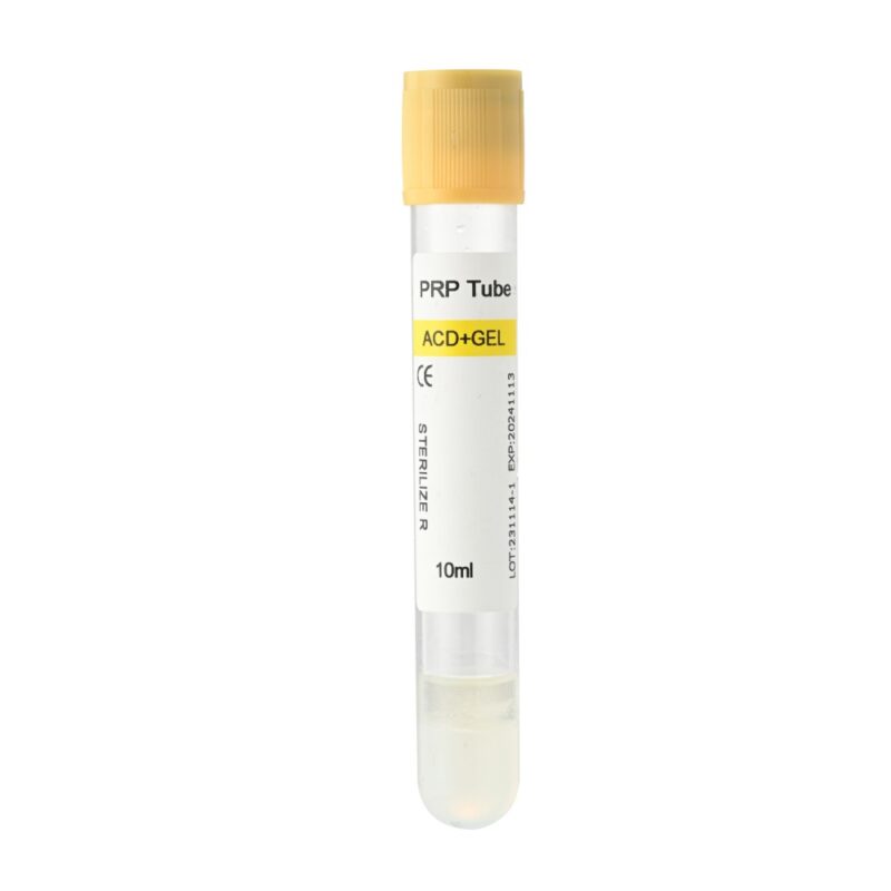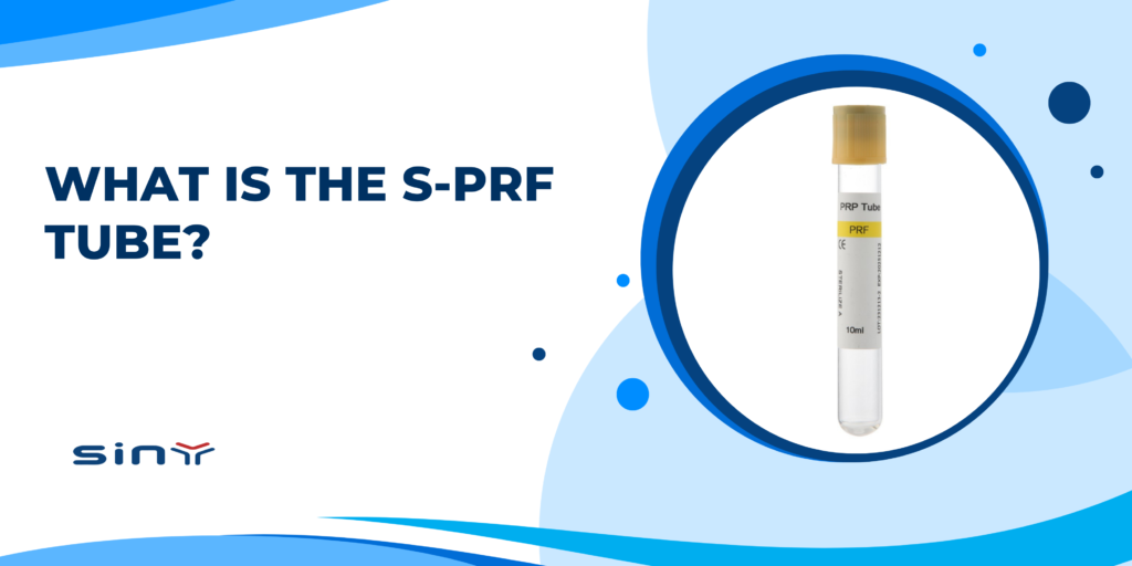
The Solid Platelet-Rich Fibrin (S-PRF) tube lets clinicians harvest platelets and growth factors from a patient’s blood in a single, solid clot. Since the late 1990s, developers have fine-tuned centrifuge settings and refined tube surfaces to create an easy-to-use device with high biological activity. Because S-PRF relies solely on the patient’s blood, with no additives, it now serves fields from dental implants to soft-tissue repair and aesthetic treatments.
Composition and Mechanism
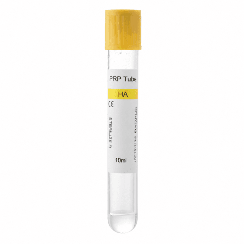
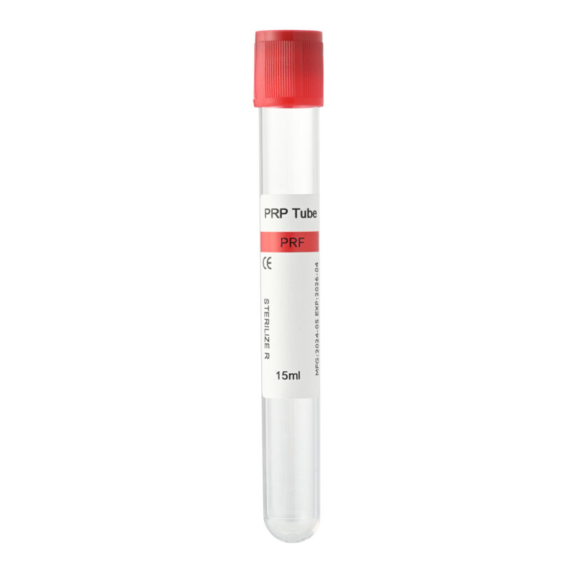
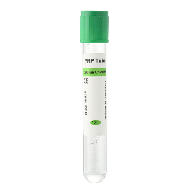
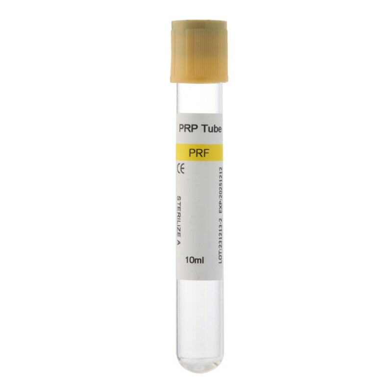
- Tube Material: We use medical-grade polypropylene (PP) or polystyrene (PS). Plasma or chemical treatment on the inner wall creates a blood-friendly surface that triggers fibrin to link into a three-dimensional network.
- Biochemical Process: With no or minimal anticoagulant, the centrifuge spin activates platelets to release PDGF, TGF-β, VEGF, and other key factors. Under shear stress, fibrin monomers self-assemble into a scaffold that traps platelets and white cells, enabling sustained factor release and a regenerative microenvironment.
Preparation Procedure
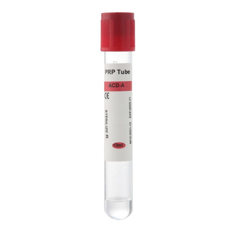
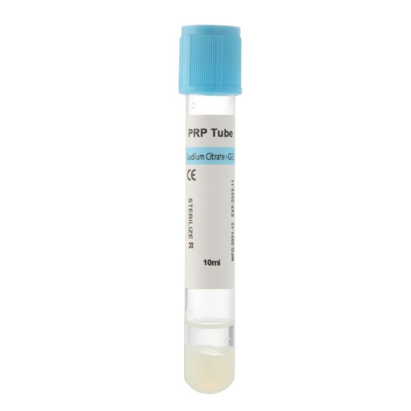
- Blood Collection: Draw 9–10 mL of venous blood without vigorous shaking.
- Centrifugation: Spin at 1,200–1,500 × g for 6–8 minutes at room temperature (18–24 °C).
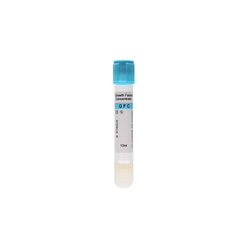
- Layer Separation: After spin-down, the tube contains three layers: red cells at the bottom, the S-PRF clot in the middle, and clear serum on top.
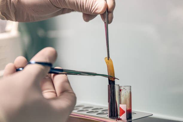
- Clot Retrieval: Use sterile forceps to lift out the middle clot, blot away excess serum, and apply it directly or mix it with bone particles or other biomaterials.
Key Quality Control Points
- Sterile Technique: Perform collection, centrifugation, and handling in a Class I or II clean area.
- Centrifuge Calibration: Check speed and time settings regularly to ensure consistent results.
- Tube Surface Verification: Choose tubes certified to international standards to guarantee reliable fibrin network formation.
Clinical Applications and Research Progress
- Dental Implants: Filling the implant site with S-PRF shortens healing and reduces inflammation after surgery.
- Soft-Tissue Regeneration: Applying S-PRF to gum recession or skin wounds boosts fibroblast growth and new blood-vessel formation.
- Orthopedic Repair: Mixing S-PRF with autologous bone graft can increase new bone volume by 20–30%.
- Aesthetic Medicine: Combining S-PRF with microneedling or injection improves collagen renewal, skin texture, and elasticity.
Comparison of Advantages
- Versus L-PRF: S-PRF uses lower spin forces to produce larger fibrin pores and prolong factor release.
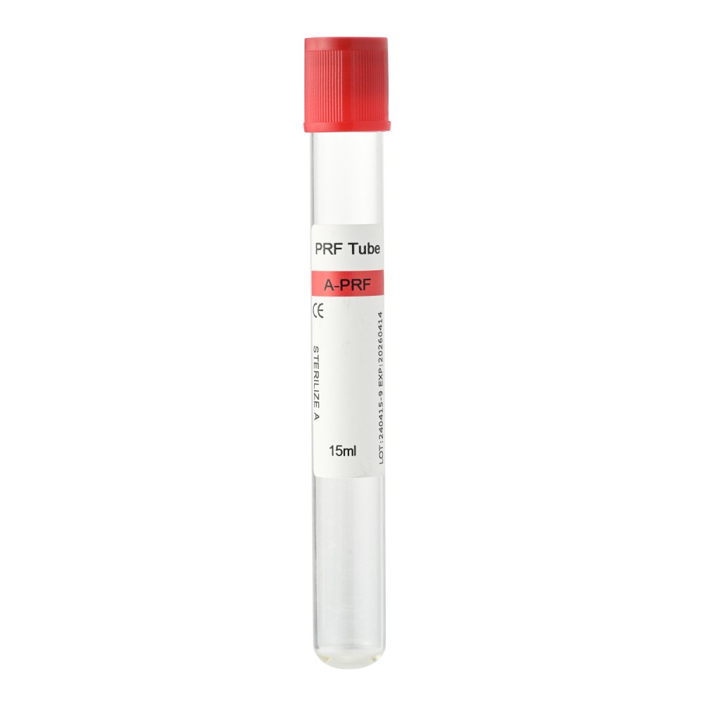
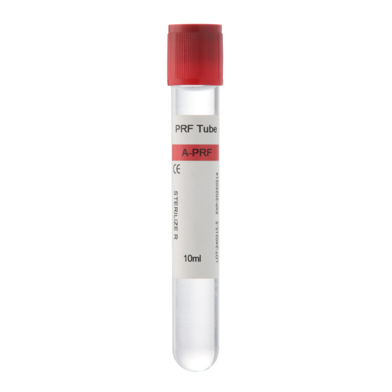
- Versus A-PRF: A-PRF relies on decreasing-speed spins that yield smaller clots. S-PRF clots remain thicker, carrying more cells and factors.
- Additive-Free: Relying entirely on the patient’s blood avoids external thrombin or high-dose anticoagulants, maximizing biocompatibility.
Usage Precautions
- Time Window: Use each S-PRF clot within 30 minutes of centrifugation to preserve factor activity.
- Custom Shaping: Trim or compress the clot as needed to fit any defect precisely.
- Temperature Control: Keep the procedure room between 18 °C and 24 °C. Cold delays clot formation; heat speeds degradation.
Summary
3By capturing a patient’s own platelets and growth factors in a three-dimensional fibrin scaffold, the S-PRF tube delivers a fast, reliable autologous repair method. Standardized protocols and rigorous quality checks guarantee consistent outcomes. Looking ahead, integrating smart centrifuges and advanced biomaterials will broaden S-PRF’s role in dentistry, orthopedics, dermatology, and aesthetic medicine, offering patients safer, more effective tissue-regeneration therapies.
FAQs
How does the S-PRF tube differ from traditional PRF?
Refining spin speed and duration yields a thicker, more uniform fibrin clot. Special tube treatments ensure steady growth-factor release.
How long can I store an S-PRF clot?
For peak potency, apply the clot within 30 minutes of centrifugation. Delays can reduce biological activity.
Can I adjust centrifuge settings?
The standard 1,200–1,500 × g for 6–8 minutes balances clot size and factor concentration. Any change requires validation against clinical goals.
What materials pair well with S-PRF?
Clinicians often combine it with autologous bone, bio-ceramics, or scaffold materials to boost bone or soft-tissue regeneration.
Which patients should avoid S-PRF?
Those with severe thrombocytopenia, active infections, or coagulation disorders should undergo hematologic evaluation before use.
How can I improve my preparation consistency?
Keep a strict sterile field, maintain room temperature, calibrate your centrifuge regularly, and use certified tubes operated by trained staff.