Tissue fixative plays a key role in preserving biological samples for medical diagnosis, research, and education. It helps keep cell structures and molecules intact, ensuring tissue details remain clear during slide preparation and subsequent analysis. This article explains the basic concepts, main components, working mechanisms, practical applications, and safety measures related to tissue fixatives. The goal is to provide medical professionals, researchers, and educators with a clear and professional reference.
Basic Concepts and Importance
Definition and Function
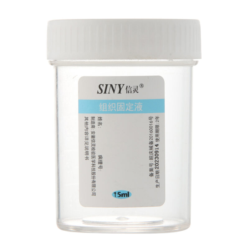
Tissue fixative is a chemical solution designed to preserve biological tissues. It locks proteins, nucleic acids, and other molecules in place. This process helps maintain the original arrangement of cells and structures so that later procedures like staining and microscopic examination yield accurate results. In effect, tissue fixative acts as a “preserver” that stops the sample from degrading.
Clinical and Research Significance
- Pathology Diagnosis: In clinical practice, proper fixation ensures that tissue samples reflect true cell morphology. This accuracy is essential for correct staining and antibody testing, which in turn supports reliable disease diagnosis.
- Scientific Research: Researchers use tissue fixatives to secure cell structures for studies such as immunohistochemistry and molecular analysis. A well-fixed sample provides valuable insights into disease mechanisms and drug effects.
- Medical Education: High-quality fixed tissue slides allow students to observe clear anatomical and pathological details. These slides help bridge the gap between theoretical knowledge and real-life observation.
Main Components and Working Mechanisms
Common Types of Fixatives
- Formalin-Based Fixatives: The most widely used solution is a 10% formalin fixative. It contains formaldehyde, which quickly penetrates the tissue and forms stable bonds with proteins. This method produces consistent results and is cost-effective, although it may sometimes mask antigen sites in certain tests.
- Alcohol-Based Fixatives: Solutions such as ethanol or methanol act by removing water from the tissue and precipitating proteins. These fixatives suit specific staining techniques and some immunohistochemical tests.
- Specialized Fixatives: Options like Bouin’s solution or zinc-based fixatives address particular experimental needs. They balance the preservation of tissue structure with the retention of molecular details, although they require careful control of conditions during use.
Working Principles
- Crosslinking Reaction: In formalin-based solutions, formaldehyde reacts with amino acids to form crosslinked bonds. This process holds cell membranes, nuclei, and organelles in place, which helps maintain the tissue’s original structure.
- Precipitation and Dehydration: Alcohol fixatives work by dehydrating tissues and causing proteins to precipitate. This method preserves overall structure while reducing cell swelling and distortion.
- Combined Effects: Some fixatives combine crosslinking with pH adjustments or ion concentration control. These formulations optimize tissue preservation for specific types of molecular tests.
Applications and Operational Practices
In Pathology Diagnosis
Tissue fixatives serve as the first step in preparing samples for diagnosis. They help preserve cellular details that pathologists rely on to detect changes and identify diseases. For instance, accurate fixation is vital in tumor diagnosis, where cell shape and nuclear details influence treatment decisions.
In Scientific Research
High-quality fixed tissues support various experimental methods:
- Immunohistochemistry: Fixatives preserve antigen structures, which allows antibodies to detect target proteins accurately.
- Molecular Analysis: By preventing tissue degradation, fixatives help maintain RNA, DNA, and protein integrity for detailed molecular studies.
- Digital Pathology: Good fixation practices yield high-quality images that serve as reliable data for computer-assisted diagnosis and research.
In Medical Education
Fixed tissue slides offer students a practical view of human anatomy and pathology. These slides help them connect theoretical concepts with real-life observations, enhancing their learning experience.
Operational Guidelines
- Time Management: Adjust fixation time based on tissue size and density. Too short a time may lead to incomplete fixation, while too long a time can hinder staining and molecular tests.
- Concentration Control: Follow standardized dilution and mixing procedures to achieve consistent results.
- Temperature and Environment: Maintain appropriate temperature and humidity. Ensure good ventilation to support optimal chemical reactions.
- Post-Fixation Procedures: After fixation, tissues undergo dehydration, clearing, and embedding. Each step requires precise control to preserve the sample’s structure.
Safety Measures and Common Challenges
Safety Practices
Tissue fixatives, such as formalin, can irritate skin and respiratory systems. To ensure safety:
- Wear Protective Gear: Use gloves, safety glasses, and masks to reduce exposure.
- Work in Well-Ventilated Areas: Use fume hoods or work in spaces with good airflow.
- Proper Storage and Disposal: Store fixatives in cool, dry places and dispose of waste according to safety guidelines.
Common Issues and Solutions
- Uneven Fixation: Large or dense tissues may not fix evenly. Cutting tissues into smaller pieces or extending fixation time can help.
- Staining Irregularities: Incorrect fixation can affect staining quality. Adjust fixative concentration and exposure time as needed.
- Antigen Masking: Excessive fixation may cover antigen sites, which interferes with antibody detection. Use antigen retrieval techniques or select alternative fixatives if necessary.
Final Thoughts
Tissue fixative forms the bridge between clinical diagnosis, research, and education. Its careful application ensures that tissue samples retain accurate structural and molecular details, which are crucial for effective diagnosis, reliable research outcomes, and clear teaching materials. As technology advances, new fixative formulations and automated systems promise to enhance the preservation process further. By understanding the principles and practices behind tissue fixation, professionals can continue to improve sample quality and drive progress in medical science.